|
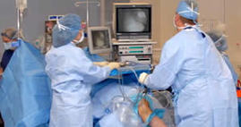
Although arthroscopic surgery has received a lot of public attention because it is used to treat well-known athletes, it is an extremely valuable tool for all orthopedic patients and is generally easier on the patient than "open" surgery. Most patients have their arthroscopic surgery as outpatients and are home several hours after the surgery. Dr. Joyce says:
"For my patients who require a simple knee scope (a debridement, meniscectomy, removal of loose bone or cartilage fragments or removal of synovial tissue) they walk out of the hospital after surgery without a brace or crutches. Most are amazed that within ten days they feel fabulous." Rehabilitation is imperative for each patient to achieve optimum recovery and range of motion. Dr. Merrill, Dr. Dukas and Dr. Joyce's patients begin a customized therapy program within days of surgery. |
|
ACL Reconstructions & Revision Surgery
The Anterior Cruciate Ligament or ACL is most commonly injured during sporting activities when an athlete suddenly pivots causing excessive rotational forces on the ligament. Individuals who experience ACL tears usually describe a feeling of the joint giving out, or buckling; people also often say they hear a “pop.” One of the initial signs of an ACL injury is pain and swelling of the joint. In order to repair the ligament, both Dr. Joyce and Dr. Dukas perform an ACL Reconstruction and use a graft, often from the patellar tendon, to reconstruct the ACL, it can’t simply by sutured back together. This is best done after initial swelling from the injury subsides. The ACL Surgery will be done arthroscopically or minimally invasive with just a few small incisions, decreasing recovery time, pain, and time in the hospital. After surgery, post-operative physical therapy is crucial to restore function and range of motion to the knee. Dr. Joyce, Dr. Dukas, and Dr. Merrill will follow your care closely to ensure our patients get back to their preinjury self.
Occasionally, people who have undergone ACL reconstructions re-tear the ACL and graft. When this happens, Dr. Joyce , Dr. Dukas, and Dr. Merrill will perform an ACL revision surgery. They both have extensive experience and adeptly skilled in handling ACL revisions. Much like the last one, the procedure will be done arthroscopically, but the graft must be harvested from a different part of the body, since the patellar tendon has most likely already been used. Often times this new graft comes from the hamstring or, occasionally, the quadriceps tendon. Sometimes the previous tunnels created during the last surgery are not amenable to a single procedure revision and will have to be bone grafted and the ACL reconstructed in a staged fashion with a second surgery. Dr. Joyce and Dr. Dukas perform a high volume of revision and primary ACL reconstructions in Connecticut and will choose which is best for the patient and, once again the patient will complete a course of physical therapy to strengthen and restore function and stability to the knee.
Occasionally, people who have undergone ACL reconstructions re-tear the ACL and graft. When this happens, Dr. Joyce , Dr. Dukas, and Dr. Merrill will perform an ACL revision surgery. They both have extensive experience and adeptly skilled in handling ACL revisions. Much like the last one, the procedure will be done arthroscopically, but the graft must be harvested from a different part of the body, since the patellar tendon has most likely already been used. Often times this new graft comes from the hamstring or, occasionally, the quadriceps tendon. Sometimes the previous tunnels created during the last surgery are not amenable to a single procedure revision and will have to be bone grafted and the ACL reconstructed in a staged fashion with a second surgery. Dr. Joyce and Dr. Dukas perform a high volume of revision and primary ACL reconstructions in Connecticut and will choose which is best for the patient and, once again the patient will complete a course of physical therapy to strengthen and restore function and stability to the knee.
Complex Multi-ligament & PCL Reconstructions
The PCL keeps the shinbone from moving backwards too far. It is stronger than ACL and is injured less often. Many times, a PCL injury occurs along with injuries to other structures in the knee such as cartilage, other ligaments, and bone. PCL injuries often happen with sudden blows to the front of the knee, twisting motions, or hyperextension injuries. When there is a complete tear to the PCL, reconstruction of the ligament is necessary to restore stability, function, and range of motion to the knee. Much like the ACL reconstruction, a graft will be used and arthroscopic surgery will be performed by Dr. Joyce. Physical therapy will happen within days of surgery and compliance is integral to the patient’s recovery.
Injuries to the collateral ligaments are usually caused by a force that pushes the knee sideways. These are often contact injuries. Medial collateral ligament (MCL) tears often occur as a result of a direct blow to the outside of the knee. This pushes the knee inwards causing pain to the inside of the knee. Blows to the inside of the knee that push the knee outwards may injure the lateral collateral ligament (LCL) causing pain to the outside of the knee. These injuries often occur in conjunction with injuries to the cruciate ligaments.
These complex multi-ligament surgeries are still done arthroscopically, but take more time to reconstruct the torn cruciate ligament, repair or resect the damaged menisci, and any other procedure Dr. Joyce needs to perform to repair the damage in the knee. These larger surgeries often involve a longer recovery period and more time in physical therapy.
Meniscal Injuries
One of the most commonly injured parts of the knee, the meniscus is a wedge-like rubbery cushion where the major bones of your leg connect. A strong stabilizing tissue, the meniscus helps the joint carry weight, glide, and turn in many directions.
Menisci tear in a number of different ways. Sudden tears often happen during sports when players squat and twist the knee. Older people, however, are more likely to have degenerative meniscus tears. Cartilage weakens and wears thin over time. These aged, worn tissues are more prone to tears. Common symptoms may include a “popping” sensation but may still be able to walk, or even keep playing, stiffness, swelling, and tenderness in the joint line, a collection of fluid in the joint slipping, popping, or locking of the knee associated with a loosened fragment.
The decision to operate or proceed with conservative treatment depends on many different factors which our providers will discuss with the patient.
Patella Dislocations
The patella, often knows as the kneecap, connects the muscles in the front of the thigh to the shinbone, or tibia. As you bend or straighten your knee, the kneecap is pulled up or down. In a normal knee, the kneecap fits nicely into the femoral groove, a V-shaped notch at one end of the femur. If the groove is uneven or too shallow, the kneecap could slide off, resulting in a partial or complete dislocation. In a chronic condition, arthroscopic surgery may be required to correct the instability. Your physician may elect to realign and tighten the tendons that keep the kneecap on track, or to release tissues that pull it off track. He will determine the best course of treatment based on an MRI and patient evaluation.

